Imagine we get access to a fourth dimension. We have access to three dimensions like we know. The fourth dimension is imagined in many sci fi movies and physics theories and it always is time. If you get access to this dimension, which is time, you can travel through time. Something very close to time travel in the field of biology or assisted reproductive technology is time lapse microscopy. Using this technology, you can go back in time and see what happened at what time and rectify the mistakes, invent solutions, and learn to tackle future problems. So today, we’ll be understanding time lapse microscopy, what this technology actually is, and how it can be applied in, assisted reproductive technology. We’ll learn embryo morphology and morphokinetics that can be identified with the help of time lapse microscopy. We’ll also learn to use, time lapse microscopy to predict blastocyst formation, implantation, and live bird potential. We’ll look into the effects of embryos, laboratory, and maternal factors on the morphokinetic parameters as observed by time lapse microscopy.
TLM technology
Digital cameras capture images of embryos at set time intervals, which can be played back as a time lapse sequence to evaluate embryo morphology and morphokinetics. Majorly morphokinetics, which is very difficult to evaluate if you don’t have TLM technology. The device used in ART setups for TLM technology or time lapse in microscopy technology is called an embryo scope. So this is majorly used in eSET cycles, which is also called elective single embryo transfer cycles.
This awards ART associated multiple birds. In these cycles, we transfer one very good embryo that has been formed so that we avoid multiple birds. Embryoscope. So this is a device used in art setups to evaluate the morphokinetics of embryos. So this is a type of incubator that allows the embryologists to monitor the morphology and morphokinetics of embryos while they are still in the incubator Embryo morphology.
The first morphological event that happens after sixteen to eighteen hours of insemination is the presence of two pronuclei. The first thing we’ll, we can use embryo scope for is for fertilization check. If we can see if the fertilization has happened properly, the TLM technology can help identify the transient pro nucleus and, avoid discarding deployed zygotes that have undergone early pronuclear disappearance or fusion of two pns to form only one pn. So we know syngamy happens after the two pronucleus are appeared. So syngamy could happen way before, like, around sixteen to eighteen hours.
It is very important for us to identify if it has two nucleus. The event is a transient occurrence. TLM can identify it with great sensitivity than single time point assessment. Multinucleation is reflective of the timing of morphokinetic parameters of developing embryos fragmentation.
What is fragmentation?
Fragmentation is basically residues of cytoplasm that are left out when, like, the cleavage happens and the extra cytoplasmic stuff that is not included inside the cell is just, left out inside the embryo. These are called fragmentation. Imagine breaking a bread and the crumbs falling down. The two breads are the two cleaved, the blastomeres, and the crumbs can be referred to as the fragments. The causes can be abnormal pH, temperature, and oxygen tension. If there is any abnormality in these parameters, the fragmentation will be high. Single time point assessment can miss the presence of the nucleus, which is disassembled during the mitotic phase. This compromises the accuracy of embryo scoring. So sometimes the, fragments could be very big. Sometimes they can be very small.
What is this blastocoel?
It is a cavity in the middle of the embryo. This for other formation of blastocoel happen and then full blastocyst formation. Now the embryo can be called a blastocyst because there is a blastocyst in the middle and the blastomeres are covered around it. Expansion of blastocyst happens after that. And then the blastocyst cavity contraction happens.
The prediction of blastocyst potential or blastocyst quality early in the embryo culture is very important part of the Clinical Embryology courses in India. And time lapse technology or time lapse, microscopy technology can help us predict implantation much earlier by looking at how the morphokinetics of the embryos have gone through. Can we predict live bird potential using an embryo scope? The ultimate goal of an IVF center is for patients to carry home a healthy live born baby. A universe set analysis of morphology showed that additional four cell embryo availability and presence of early cleavage significantly increases odds of live birth.
Aneuploidy is a common occurrence in human embryos with estimates ranging from twenty percent to ninety percent depending on the woman’s age. The only current existing method to identify aneuploidy embryos prior to transfer involves invasive embryo biopsy with genetic screening. We only can do PGT or, pre implantation genetics to know if the aneuploidy is present or not. Existing data regarding the ability of, time lapse microscopy and morphogenetic analysis to detect aneuploidy embryos are conflicting. But yes, there are like studies that say that, yes, you can tell if there are there is an aneuploidy present in this embryo to an extent.
Increasing maternal age is that generally associated with decreasing oocyte quality, increased risk of aneuploidy and decreased embryo quality. The maternal age does not seem to have an effect on embryo developmental kinetics. Age does not have much effect on the embryo morphokinetics.
Obesity has been shown to be correlated with lower implantation and pregnancy rates. The effect of obesity on embryo kinetics is currently inconclusive. So a lot of research is open in this area. Similar to obesity, the effect of PCOS is embryo kinetics is also currently inconclusive. We don’t know if BMI PCOS or age can have a direct effect on the morphokinetics of the embryo.
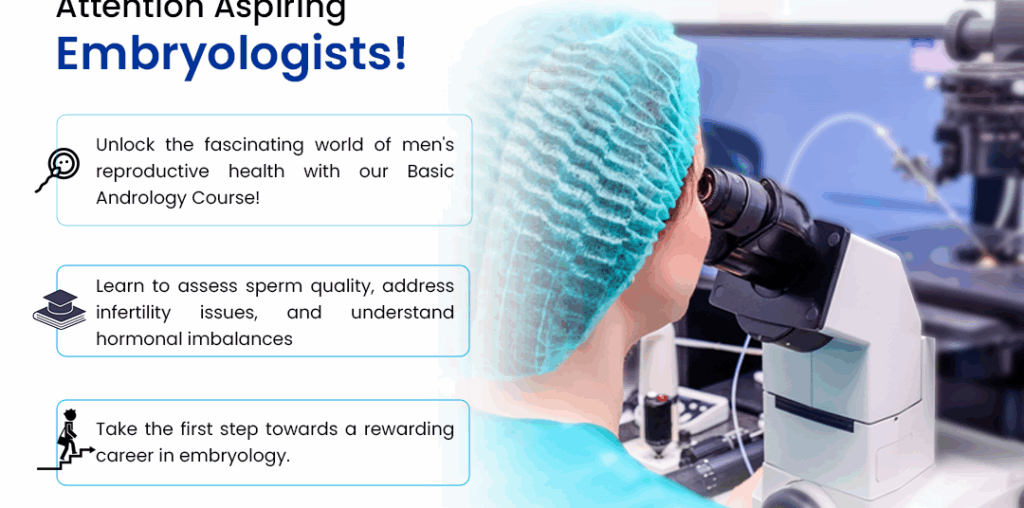
Embryology Courses in Bangalore
Commenced under the leadership of Padma Shri Prof Dr. Kamini A Rao, Medline Academics recognizes the immense potential of Embryology courses in Bangalore as a promising career path for M.Sc. students. With a well-structured curriculum, state-of-the-art laboratories, research opportunities, expert faculty, workshops, and placement support, Medline Academics is dedicated to preparing aspiring embryologists for successful careers in the field. Medline Academics ensures that the faculty members teaching the Clinical Embryology Courses in India program are highly accomplished and experienced in their respective fields. The expertise of these faculty members enriches the learning experience and offers valuable insights into industry best practices. Medline Academics provides career-oriented support to its students. The institution assists graduates in finding suitable job opportunities in the field of clinical embryology, ensuring a smooth transition from academia to a successful professional career. Through its holistic approach to education, Medline Academics prepares students to become competent and responsible clinical embryologists, making a positive impact on the field of assisted reproductive technology and reproductive medicine.
Looking for an IVF specialist in Bangalore?
Come to Dr. Kamini Rao Hospitals, where you get the best treatment for fertility. This hospital with 2 centres in Kumarapark and Jayanagar, has the best IVF Specialists in Bangalore. Besides ART Procedures like IUI, IVF and ICSI, the hospital also has wide range of services like general gynaecological, menopause, Counselling etc. Besides, patient care, this hospital also trains mentees for Fellowship in Reproductive Medicine in India and Fellowship in Embryology. They don’t just observe, they also assist the Sr. Doctors for performing complex procedures.